Part A Drag the correct label to the appropriate location to identify the structures of a synovial joint. Solution for 3D Ligament of the femaral head.
Art Labeling Activity Anatomical Structure Of The Chegg Com
Angular Movements of the Joints Learning Goal.
. Structure of a Synovial Joint Label the structures of a synovial joint. Chapter Test - Chapter 8 Question 19 The articular cartilage damaged by osteoarthritis is characterized by. Bone Markings on the Right Femur anterior surface Bone Videos.
Identify and test your knowledge of the locations of the muscles of the torso with this great free quiz - Anterior locations Quiz 2. Introduction to Anatomy and Physiology 1st Edition Michelle Provost-Craig Susan J. The hip joint is one of the most important joints in the human body.
The right elbow joint medial view PICTURE. Lecture Exam 1- AP Semester 1. Joint movements flexion and extension Recommended textbook explanations.
Bones of the Right Wrist and Hand anterior view Art-labeling Activity. With the hip flexed the range of. The right knee joint anterior view superficial layer Art-labeling Activity.
Learn vocabulary terms and more with flashcards games and other study tools. Factors that increase the stability of the hip joint include. Use the art-labeling activities to quiz yourself on key anatomical structures in this chapter.
Weve got the study and writing resources you need for your assignmentsStart exploring. Start studying Art-labeling Activity. The structure of a long bone humerus of arm Figure 59.
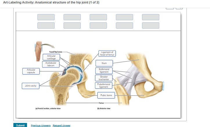
The right hip joint. Anatomical structure of the hip joint 1 of 2 Ligament of head of femur Rp bones Articular cartilage Acetabular laborum llium Articular capsule lofemoral ligament Greater trochanter Joint cavity Pubofemoral ligament Pubic bone Fermer Whos section. Correct Help Reset Help Reset Medullary cavity Spongy bone Periosteum Fibrous joint capsule Synovial.
The pectineus and iliopsoas muscles are responsible for movement at the hip and are discussed elsewhere. The Right Humerus anterior and posterior surfaces Art-labeling Activity. Grading Policy Art-labeling Activity.
Burning through metal and st. A a rough network of bristly collagen fibers. The Knee Joint Drag the correct label to the appropriate structure of the knee joint.
1059pm on Sunday February 7 2021 You will receive no credit for items you complete after the assignment is due. Rotational Movements of the Joints. It allows us to walk run and jump.
This forms an immobile synarthrosis type of joint. The Right Hip Joint Art Labeling Activity Anatomical Structure Of With the hip flexed the range of. Anatomical structure of the hip joint 1 of 2 Abutabular forum Tobolomical ligament Ligament of head of femur Articular cartilage Articular capsuide Joint cavity llofemoral ligamont Pubic bone Grade trochanter Pued hip bones Femur hotel con anteriori Arterie.
Deadly draw nadder wallpaper. When the two hip bones are combined with the sacrum and coccyx of the axial skeleton they are referred to as the pelvisThe right and left hip bones also converge anteriorly to attach to each other at the pubic symphysis. Step 3 Turn the center v shape into a heart shape.
Yet the hip joint is also one of our most flexible joints and allows a greater range of motion than all other joints in the body except for the shoulder. The pubic symphysis is a slightly mobile amphiarthrosis cartilaginous joint where the pubic portions of the right and left hip bones. The right hip joint.
Chapter 73 Anatomy module. Human skull inferior view mandible removed. Anatomy and Physiology questions and answers.
Human skull superior view top of cranium removed Figure 511. Originates from the pelvis and attaches to the tibia. Reset Help Fibula Lateral meniscus Anterior cruciate ligament Ligaments That Stabilize the Knee Joint Tbial collateral ligament Articular cartilage Fibular collateral ligament Patellar surface Tibia Patelar igament cut Posterior cruciate ligament Medial meniscus.
Week 2 Chapter 8_ Due. The right elbow joint latera. Bio 165 Chapter 2.
Wrist is composed of carpal bones. It bears our bodys weight and the force of the strong muscles of the hip and leg. Part A Drag the labels onto the diagram to identify the angular movements of the joints.
Flexing of the lower leg at the knee joint. Anatomy and Physiology questions and answers. Pictures of joint movements.
To learn the angular movements of the joints. The right elbow joint lateral view PICTURE. How to draw a deadly nadder.
Parts of the Right Scapula Learning Goal. To see a muscular system diagram from the posterior back view click here. This muscular system picture shows all the major muscle groups on the human body from the frontal view.
Human skull lateral view. Label the angular movements of the joints. Learn vocabulary terms and more with flashcards games and other study tools.
The right elbow joint lateral view Sets found in the same folder. If a knee or hip. The structure of a Written By wilfordbellhouse35273 April 22 2022 Add Comment Edit.
Activity hip the wallpaper. Help Reset Femur Fibula Metatarsal bones Pelvic girdle Phalanges Hip bone Lower limbs Tarsal bones Tibia Patella. To learn the parts of the scapula.
The sartorius a thin muscle in the thigh the is the bodys longest muscle. The first sternocostal joint is a synchondrosis type of cartilaginous joint in which hyaline cartilage unites the first rib to the manubrium of the sternum. The two hip bones also called coxal bones or os coxae are together called the pelvic girdle hip girdle and serve as the attachment point for each lower limb.
Label the parts of the scapula. Attached to the rest of the skull by a freely movable joint. Start studying Art-labeling Activity.
Angular Movements of the Joints. Art Labeling Activity. Part A Drag the labels onto the diagram to identify the parts of the scapula.
Ditulis Salvador Owens Selasa 10 Mei 2022 Tulis Komentar Edit.

Pin By Anna Ewert Pittman On Taping Knee Joint Anatomy Human Knee Joints Anatomy
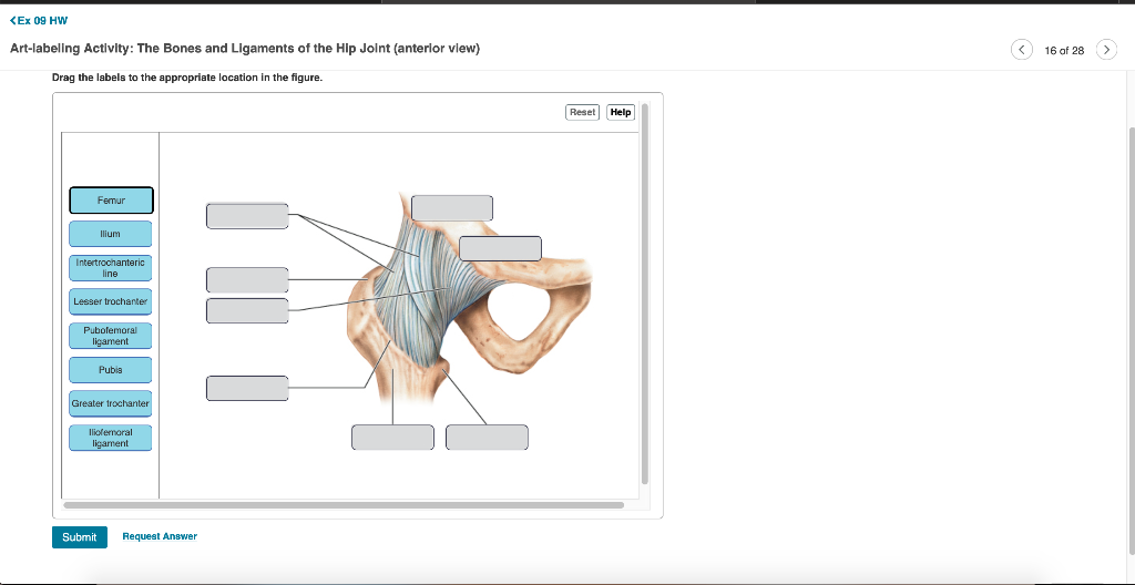
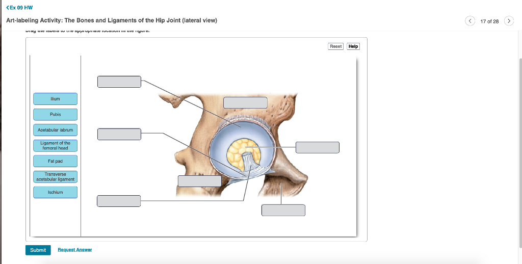
Solved Ex 09 Hw Art Labelling Activity The Bones And Chegg Com
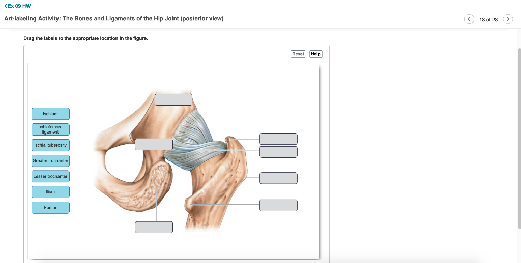
Solved Ex 09 Hw Art Labelling Activity The Bones And Chegg Com

Radiology Quiz 44015 Radiopaedia Orgviewing Playlist Rotorua Teaching Hip And Pelvis Radiopaedia Org

A P Lab 1 Practical 2 Flashcards Quizlet
Solved Ex 09 Hw Art Labelling Activity The Bones And Chegg Com
Hip Joint Illustrations Image Radiopaedia Org
2
0 comments
Post a Comment